/filters:quality(70)/prod01/prodbucket01/media/durham-university/research-/new-research-and-business/Research-and-Business-Partnerships.png)
/filters:quality(70)/prod01/prodbucket01/media/durham-university/research-/research-institutes/biophysical-sciences-institute/Screenshot_6-2-2026_95843_stories.durham.ac.uk.jpeg)
Art Inspired by Science Exhibition
This exhibition displayed scientific images of biological systems alongside creative responses from professional artists and the wider public. The exhibition explored the intriguing parallels between exploration and creativity in science and art.
About the Biophysical Sciences Institute
The exhibition has been brought together by Durham University’s Biophysical Sciences Institute. We are a Durham University Research Institute whose community includes over 150 academics and researchers from across 7 departments. Our members are united by an innovative, interdisciplinary approach to the research of fundamental biological processes which drives new discoveries and technologies.
Find out more: About Us
About the Exhibition
Through 2024-2025 creative responses were invited in response to a series of scientific images collected by Dr Robert Banks during his career teaching and researching in Durham.
More information about the scientific images can be found here.
The creative responses were curated to form our Botanic Garden exhibition and as an online exhibition. Further information about the scientific images, the exhibits and the creatives who have taken part can be found below.
Creative Responses
-
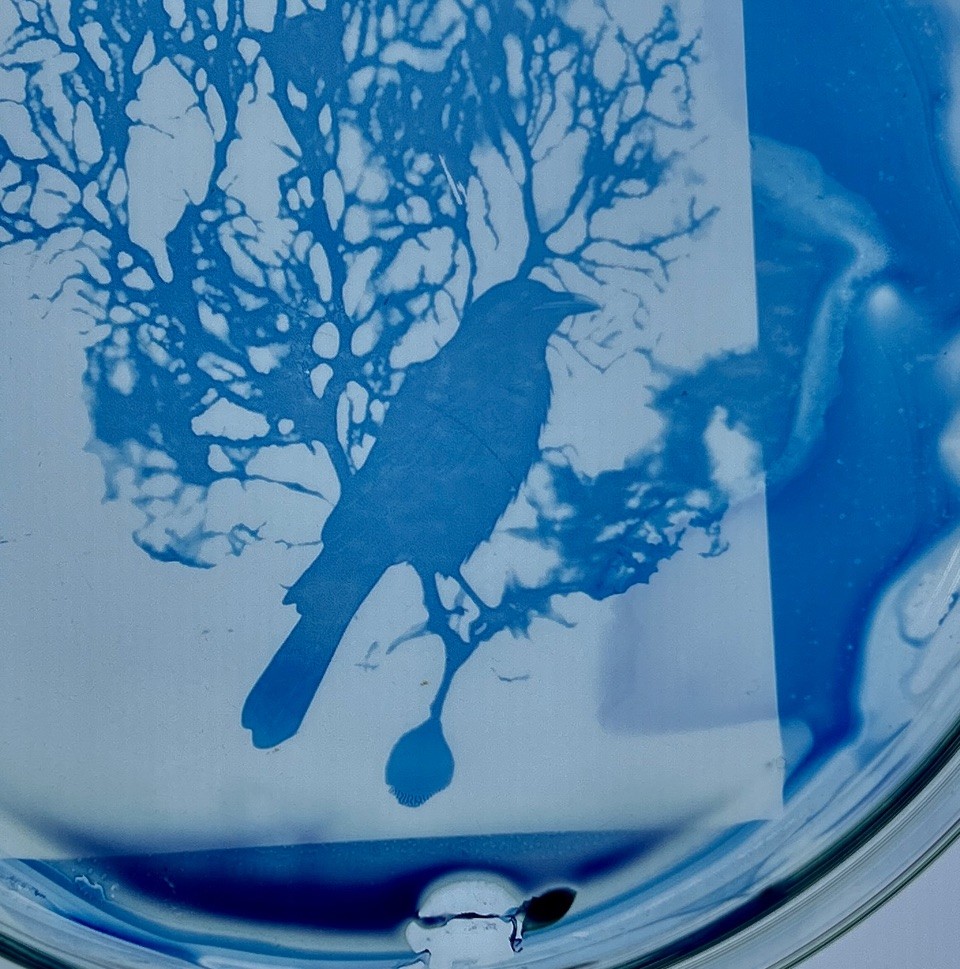
Crow & Tree (or Dust of Snow by Robert Frost)
Marianne Wilde
-
The Tree of Life
Juliet Williams
-
Curved Straight Lines
Harrison Drummond
-
We Are One
Judith Hurst
-
i
Maggie Parker
-
Cyanotype Fabric Collage
Frankie Wallace
/filters:quality(70)/prod01/prodbucket01/media/durham-university/research-/research-institutes/biophysical-sciences-institute/biophysical-stock-images/thumbnail_Crow--Tree-Petri-Dish-Detail.jpg)
Crow & Tree (or Dust of Snow by Robert Frost)
The Tree of Life
/filters:quality(70)/prod01/prodbucket01/media/durham-university/research-/research-institutes/biophysical-sciences-institute/biophysical-stock-images/20250327_132957.jpg)
Curved Straight Lines
/filters:quality(70)/prod01/prodbucket01/media/durham-university/research-/research-institutes/biophysical-sciences-institute/biophysical-stock-images/thumbnail_IMG_4148_Harrison.jpg)
We Are One
/filters:quality(70)/prod01/prodbucket01/media/durham-university/research-/research-institutes/biophysical-sciences-institute/biophysical-stock-images/We-Are-One_JH_crop.jpg)
i
/filters:quality(70)/prod01/prodbucket01/media/durham-university/research-/research-institutes/biophysical-sciences-institute/biophysical-stock-images/thumbnail_image_05_MP_PNG-294X379.png)
Cyanotype Fabric Collage
/filters:quality(70)/prod01/prodbucket01/media/durham-university/research-/research-institutes/biophysical-sciences-institute/biophysical-stock-images/thumbnail_image1.jpg)
Further Information
Dance of the Cells
Charlotte Bassadone
The Dance of the Cells project is a response to an image of the gastro glands lining the stomach, collected by Dr Robert Banks during his career in comparative neuroscience, and in particular exploring the structures that act at the junction between muscle and nerve cells. The images capture a moment in time, showing the delicate interplay between pale and darker cells, secretion and the process of cell division. By translating these microscopic forms into four watercolour paintings, which themselves are at varied magnifications, Charlotte highlights the hidden elegance within our own bodies and emphasises the cell fragility and intricate layering. These are further enriched by the use of aqueous toluidine dye—echoing techniques used in light microscopy.
Watercolour, oil pastel and aqueous toluidine dye on watercolour paper
Microcosms
Sarah Calavera
Microcosms is a body of work that explores the intricate relationship between art and science, specifically focusing on the microcosmic worlds within the human body. The title reflects this concept: while the human body itself is often considered a microcosm, it is made up of countless little worlds—cells, nerves, and fibers—that create complex, interconnected systems. Each of these elements forms its own distinct, yet integral, universe within us.
Microcosm #1
Sarah Calavera 2025
Mixed media on canvas, Xuan rice paper, Gelli print, floral netting, text from Gray’s Anatomy book pages, assorted papers, distress inks
Microcosm #2
Sarah Calavera
Triptych 3 Wood Panels
Mixed Media on wood panels, collage of vintage anatomy illustrations, floral netting, Xuan rice paper, distress inks
Curved Straight Lines
Harrison Drummond (8 years old)
Harrison carefully measured with a ruler and drew shapes with straight lines in a repeating pattern. He noticed the effect that some of the lines begin to look curved. Using straight lines and a ruler, the artist meticulously measured where the lines spread out from a central square.
He was interested that the result is that the lines appear curved. Photocopies of the image were fitted around the drawing, creating an effect he enjoyed. Harrison’s grandma reflected that there are no truly straight lines anywhere; they are all slightly curved. Harrison believes his construction reflects this idea.
Pen on paper, photocopy
Semiosomatic
Martha Gray
'Semiosomatic’ is a blending of the two words semio (from semiotics, the study of signs and symbols) and somatic (relating to the body). Martha is an artist living with chronic health issues that affect both her muscular and nervous systems, as a result she is interested in how these systems can develop communication dysfunction overtime in the form of chronic pain. Her work is a response to Dr Banks’s research into ‘the area of comparative neuroscience exploring the structures that act at the junction between muscle and nerve cells’ through the lens of her personal experience.
Bioplastic cyanotype print on glass
Particulate Tadpoles in a Quantum Pond, Patterns in a Quantum Pond & We are One
Judith Hurst
Because in real life cell structures are 3 dimensional, Judith represented this quality with layers of paper, upon which the cell structures spiral outwards from the lowest surface, morphing upwards to the higher ones.
A plaited Celtic design links everything together- forming life.
Life is seen here as flying insects, which also move in 3 dimensions.
But why a snail too?
She considers time to be the 4th dimension.
Judith believes that a snail may perceive faster life forms, such as humans, appear transitory- quick flashes of movement outside a snail’s comprehension, perhaps.
Therefore, time is relative to each life form. A bit like cells- pulsing and changing according to environment and sometimes, chance.
Vellum (calfskin), oil paint, ink, 24ct gold leaf, 18ct gold wire and hallmarked button, handmade oak frame
i
Maggie Parker
Eco-printing is a contemporary application of the traditions of natural dyeing. In eco-printing, plants are enclosed in paper, stacked in layers and then steamed or immersed in hot water to extract the pigments and produce a print made with plant dyes. Direct and close contact between the plant and the substrate is essential. Leaves, stems, flowers, buds, seeds and roots may be used; also bark and wood. At different seasons of the year, different pigments may concentrate in various plant parts so great colour variability is possible.
Inhabiting a studio space in Ushaw Historic Buildings and Gardens has inspired Maggie Parker’s art practice to continue to grow. Being constantly surrounded by creative people and the gardens and woodland areas has allowed the interaction with nature which is inherent within her art practice. She utilises the method of eco-printing to appreciate this beauty.
Eco-print on Watercolour Paper
Cyanotype Fabric Collage
Frankie Wallace
Images taken from Dr Robert Banks’ micrographs used in his teaching and research at Durham University. Developed as cyanotypes, hand painted onto cotton and hand stitched.
100% cotton
Crow & Tree (or Dust of Snow by Robert Frost)
Marianne Wilde
Base ‘Tree’ Image – A nerve cell or neuron, from the cortex of the cerebellum by Robert Banks Cyanotypes were first introduced by the astronomer, scientist, and botanist John Herschel in 1842 and botanist and photographer Anna Atkins was the first to use the cyanotype to create a photographic album of algae specimens in 1843. The process involves combining ferric ammonium citrate and potassium ferricyanide to make an iron-rich sensitiser solution. As the sensitiser chemicals react to light when exposed, the coating process takes place in dim light. These chemicals are then exposed to UV light such as sunlight, which creates ferric ferrocyanide, also known as Prussian Blue.
Dust of Snow by Robert Frost circa 1923
The way a crow
Shook down on me
The dust of snow
From a hemlock tree
Has given my heart
A change of mood
And saved some part
Of a day I had rued.
Work inspired by the idea that small things matter…
Cyanotype on glass Petri Dish – Cyanotype Emulsion/Photographic Gela n/20cm diameter Petri Dish
The Tree of Life
Juliet Williams
Created in response to Image 8: a Purkinje cell. The same cell that Santiago Ramon y Cajal immortalised in his ink drawing a hundred and twenty five years ago. Juliet responded to the dendritic repetitions in form and shape in nature at all scales; trees’ root and branch systems, the lungs, river systems.
Mistaken by a friend of the artist as the Tree of Life, the title was adopted as an apt title given the Purkinje cell’s function.
Pewter on Corten Steel
Scientific Images
-
Paramecium
Paramecium
-
Outer layer of the cerebellar cortex
Outer layer of the cerebellar cortex
-
The outer layer of the cerebellar cortex
The outer layer of the cerebellar cortex
-
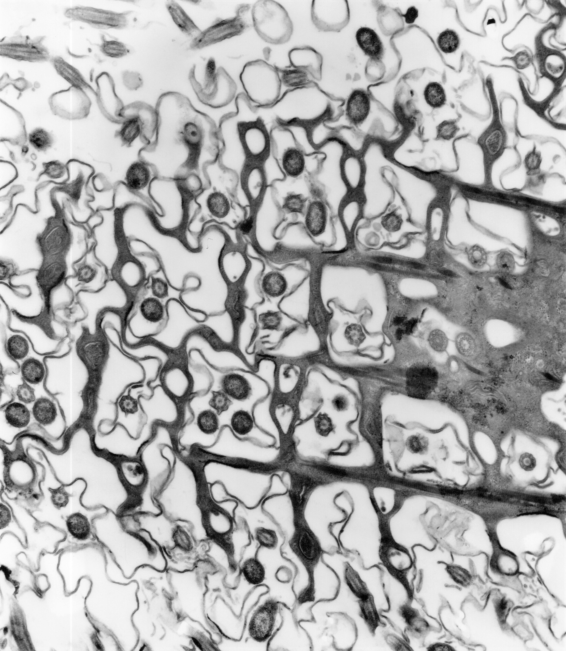
Structures Deep Within Skeletal Muscle Fibre
Structures Deep Within Skeletal Muscle Fibre
-
A nerve cell from the cortex of the cerebellum
A nerve cell from the cortex of the cerebellum
/filters:quality(70)/prod01/prodbucket01/media/durham-university/research-/research-institutes/biophysical-sciences-institute/biophysical-stock-images/paramecium05.jpg)
/filters:quality(70)/prod01/prodbucket01/media/durham-university/research-/research-institutes/biophysical-sciences-institute/biophysical-stock-images/as10.jpg)
Paramecium
Outer layer of the cerebellar cortex
/filters:quality(70)/prod01/prodbucket01/media/durham-university/research-/research-institutes/biophysical-sciences-institute/biophysical-stock-images/rat-cerebellum4434009a.jpg)
The outer layer of the cerebellar cortex
/filters:quality(70)/prod01/prodbucket01/media/durham-university/research-/research-institutes/biophysical-sciences-institute/biophysical-stock-images/mouse-mass-roi01-overview.jpg)
Structures Deep Within Skeletal Muscle Fibre
/filters:quality(70)/prod01/prodbucket01/media/durham-university/research-/research-institutes/biophysical-sciences-institute/biophysical-stock-images/em3907.jpg)
A nerve cell from the cortex of the cerebellum
/filters:quality(70)/prod01/prodbucket01/media/durham-university/research-/research-institutes/biophysical-sciences-institute/biophysical-stock-images/rat-cerebellum-01012.jpg)