

Staff profile
Dr Jacqueline Tabler
Assistant Professor
| Affiliation |
|---|
| Assistant Professor in the Department of Biosciences |
Biography
Jacqueline Tabler completed her PhD with Jeremy Green at King's College London, where she investigated how cell polarity regulates cell differentiation during early development in Xenopus. She then transitioned into craniofacial biology during postdoctoral research conducted between the laboratories of Karen Liu at King's College London and John Wallingford at the University of Texas at Austin. There, she discovered that impaired expansion of the embryonic neural crest underlies several craniofacial features of ciliopathies, including high-arched palate, laryngeal dysgenesis, and vocalisation defects.
As an independent Group Leader at the Max Planck Institute of Molecular Cell Biology and Genetics in Dresden, Jacqueline combined advanced live and fixed tissue imaging with quantitative and theoretical approaches to investigate how mechanical forces regulate tissue morphogenesis and cell fate decisions in the developing mammalian skull. This work demonstrated how reciprocal interactions between differentiating cells and the extracellular matrix coordinate tissue organization and cell fate during development.
At Durham University, Jacqueline builds on these discoveries to investigate how collective cellular behaviours emerge through reciprocal interactions between tissue geometry, mechanics, and molecular dynamics, using embryonic development as an experimentally tractable system to uncover general biological principles. Her laboratory focuses on how connective tissue layers are patterned, how stem cells make cell fate decisions, and how changes in tissue mechanics contribute to connective tissue disorders. By modelling defects in collagen crosslinking, including disruption of the enzyme lysyl oxidase, her research aims to bridge fundamental cell biology with human diseases such as Ehlers-Danlos syndrome and progeria.
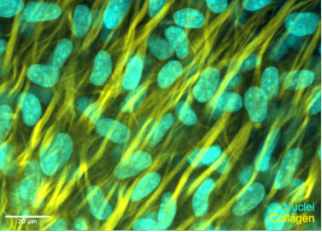
Image: Spinning disc confocal microscopy of differentiating bone mesenchyme in the embryonic mouse skull cap (E15.5) in flatmount preparation. Collagen is stained with FastGreen (Yellow) and nuclei are stained with DAPI (cyan). Scalebar, 20µm.
Publications
Journal Article
- Self-propagating wave drives morphogenesis of skull bones in vivoDang, Y., Lattner, J., Lahola-Chomiak, A. A., Afonso, D. A., Ulbricht, E., Taubenberger, A., Rulands, S., & Tabler, J. M. (2025). Self-propagating wave drives morphogenesis of skull bones in vivo. Nature Communications, 16(1), Article 4330. https://doi.org/10.1038/s41467-025-59164-9